Open Academy navigation
Gandamala and Apachi - Nidana, Samprapti, Lakshana and Chikitsa.
चयप्रकर्षात् अपचीम् वदन्ति - Named for progressive accumulation
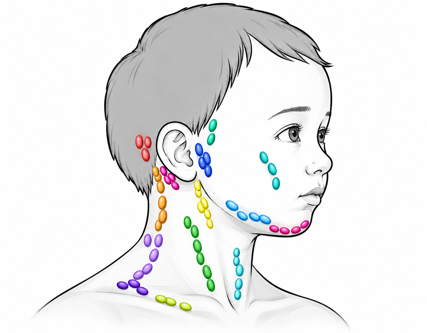 दोष-धातु:
 - भोज: त्रिदोष-मेदः
- हन्वस्थि - Jaw region
- कक्षा - Axilla
- अक्षका - Clavicular region
- बाहुसन्धि - Arm joint
- मन्या - Neck
- गल - Throat
- A/c to भोज : Starts at जङ्घाकण्डराs, प्रकुपित वात causes ऊर्ध्वगति (upward spread) to वक्षः–कक्षा–मन्या–गल
- स्थिर - Firm/fixed
- वृत्त or आयत - Round/ Elongated
- स्निग्ध - Smooth
- बहुग्रन्थि - Multiple swellings
- अनन्यवर्ण - Not different from normal skin colour
- चयप्रकर्ष - Progressive accumulation
When अल्परुज & कण्डूयुक्त - (less painful & Itchy)
- प्रभिन्ने स्रवन्ति नश्यन्ति- Breaks open - Discharge flows out - Subsides
- अन्ये भवन्ति - New swellings develop
- आमलकास्थिमात्र - Size of an आमलकी seed
- मत्स्याण्डजालप्रतिम - Fish-egg-like appearance
TYPES - दोषानुसार प्रकार
कफज - गूढ, अपाक, कठिन, स्निग्ध, अल्परुक्कर
मेदोज - same as कफज +अतिमार्दव,
If वर्षगणानुबन्धी (if persistent for years)
- सुदुस्तरा - Very difficult to treat
- A/c to भोज- व्यामिश्रदोषजा - कृच्छ्रसाध्या
अपची Vs गण्डमाला
तां तु मालाकृतिं विद्यात् कण्ठहृद्धनुसन्धिषु ।गण्डमालां विजानीयादपचीतुल्यलक्षणाम् ।।
When appearing at कण्ठ-हृद्-हनुसन्धि in a मालाकृति, the presentation is known as गण्डमाला However, the lakshanas are same as अपची.
- जीमूतक, कोशवती fruits + दन्ती, द्रवन्ती, त्रिवृत् roots → prepared as सर्पि
- Described as उदारवीर्य - highly potent.
- द्विधाप्रवृत्त - interpreted as acting in both directions / both sites.
- Indicated in प्रवृद्ध अपची - advanced Apachi.
- जीमूतक with निर्गुण्डी + जाती + बरिहिष्ठ (बालक)
- Combined with माक्षिक + सैन्धव.
- Used as a strong वमनद्रव्य.
- Especially indicated in दुष्ट अपची - severe/complicated Apachi.
- कैडर्य + बिम्बी + करवीर - processed into तैल.
- Indicated for मूर्धविरेचन - therapeutic cleansing through the head/nasal route.
- शाखोटक processed with its स्वरस → medicated तैल.
- Indicated for नस्यविरेचन - nasal cleansing therapy.
- मधूकसार - useful for अवपीड नस्य.
- शिग्रु फल or खरमञ्जरी (अपामार्ग) - also indicated for अवपीड नस्य.
- In persistent disease, मत्स्याण्डसदृश मेदो-जाल is surgically exposed and removed.
- Site described in the जङ्घा, with इन्द्रबस्ति मर्म carefully avoided.
- After removal of the fatty network, अग्निकर्म is performed to destroy the residual tissue/root and prevent recurrence.
- मणिबन्धोपरि - above the wrist
- त्रि-रेखा - three incisions
- अङ्गुल्यन्तरित - one-finger-width apart
- Intended for अपची निवृत्ति - resolution of the swelling.
क्षार - Cauterising Applications
- During the चूर्णकाल / healing stage, मसी prepared from प्रचलाक, काक, गोधा, अहि, कूर्म
- Applied along with इङ्गुदी तैल.
- श्लीपद-indicated oils are also mentioned for use.
- विरेचन - therapeutic purgation
- धूम - medicated smoking therapy
- नित्यं यव–मुद्ग भोजन - regular diet based on यव (barley) and मुद्ग (green gram).
Lymphadenopathy refers to lymph nodes that are abnormal in size and/or consistency. It is a clinical finding rather than a diagnosis.
Lymphadenopathy may be classified as:
- Localized – one nodal region involved.
- Generalized – ≥2 non-contiguous nodal regions involved, suggesting a systemic process.
- Acute – generally ≤2 weeks.
- Subacute – approximately 2–6 weeks.
- Chronic – >6 weeks.
- no single universal size threshold applicable to every nodal basin. A
- commonly used clinical definition is >1 cm,
- normal size varies by location and age. For example, inguinal nodes may normally be larger, while supraclavicular nodes are considered abnormal when palpable in many clinical contexts.
Lymphadenopathy Vs Lymphadenitis
- Lymphadenopathy: abnormal node.
- Lymphadenitis: lymph-node inflammation, usually associated with tenderness, erythema, warmth and sometimes suppuration.
- Epstein–Barr virus - infectious mononucleosis
- Cytomegalovirus
- HIV
- Adenovirus
- Influenza and other respiratory viruses
- Measles
- Rubella
- Mumps
- Herpes viruses
- Staphylococcus aureus
- Streptococcus pyogenes
- Tuberculosis
- Atypical/non-tuberculous mycobacteria
- Bartonella henselae - cat-scratch disease
- Syphilis
- Brucellosis
- Tularemia
- Histoplasmosis
- Coccidioidomycosis
- Toxoplasmosis
- Other opportunistic infections.
The infectious differential depends strongly on the geographical setting, exposure history and nodal location.
Hematological
- Hodgkin lymphoma
- Non-Hodgkin lymphoma
- Leukemia
Metastatic malignancy
- Head and neck squamous-cell carcinoma
- Thyroid carcinoma
- Breast carcinoma
- Lung carcinoma
- Gastrointestinal and other solid malignancies.
Generalized lymphadenopathy is particularly important in lymphoproliferative disorders, whereas localized lymphadenopathy may represent metastatic disease from the drainage territory.
- Systemic lupus erythematosus
- Rheumatoid arthritis
- Sjögren syndrome
- Dermatomyositis
- Sarcoidosis
- Other systemic inflammatory disorders.
Examples include:
- Phenytoin and other anticonvulsants
- Some antibiotics
- Allopurinol
- Certain antihypertensive drugs
- Drug hypersensitivity syndromes.
A careful medication history is therefore part of the evaluation.
The location of an enlarged node is one of the most useful clues to its cause, because lymphatic drainage follows relatively predictable anatomical pathways.
Region | Important nodal groups |
Head & neck | Submental, submandibular, facial/buccal, parotid/preauricular, retroauricular/mastoid, occipital, retropharyngeal |
Cervical | Anterior cervical, deep jugular, posterior cervical/posterior triangle |
Supraclavicular | Right and left supraclavicular |
Axilla | Pectoral/anterior, subscapular/posterior, humeral/lateral, central, apical |
Epitrochlear | Medial arm, proximal to elbow |
Thorax | Mediastinal, hilar |
Abdomen | Para-aortic, mesenteric, iliac |
Pelvis | External iliac, internal iliac, obturator |
Groin | Superficial and deep inguinal |
Lower limb | Popliteal |
A complete lymph-node examination should therefore include cervical, supraclavicular, axillary, epitrochlear and inguinal regions, along with assessment of liver and spleen when generalized disease is suspected.
Nodal location | Drainage/source |
Submental (IA) | Chin, lower lip, anterior floor of mouth, tongue tip |
Submandibular (IB) | Oral cavity, teeth, anterior face |
Upper jugular (II) | Tonsil, pharynx, nasopharynx, oral cavity |
Middle jugular (III) | Oropharynx, hypopharynx, larynx |
Lower jugular (IV) | Hypopharynx, cervical oesophagus, larynx, thyroid |
Posterior triangle (V) | Nasopharynx/oropharynx; thyroid and posterior neck structures |
Central compartment (VI) | Thyroid, larynx, trachea, cervical oesophagus |
Preauricular/parotid | Scalp, eyelids, conjunctiva, lateral face |
Occipital | Posterior scalp |
Retroauricular | Scalp/auricular region |
Supraclavicular | Thoracic/abdominal drainage; important metastatic site |
AAO-HNS/AJCC anatomical classification.
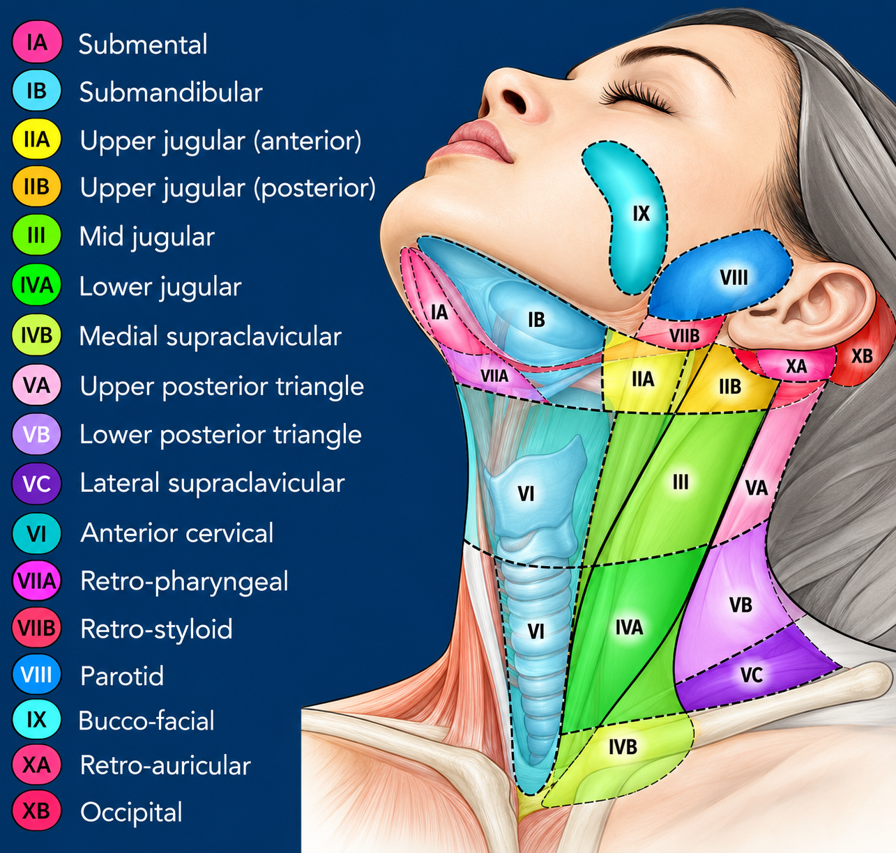 Level I
Sublevel | Group | Location |
IA | Submental | Between the anterior bellies of the digastric muscles and above the hyoid |
IB | Submandibular | Submandibular triangle, around the submandibular gland |
Major drainage: oral cavity, floor of mouth, anterior tongue, lower lip, chin and anterior facial structures.
Located along the upper internal jugular chain, from skull base to approximately the inferior border of the hyoid.
Sublevel | Location |
IIA | Anterior/medial to the spinal accessory nerve |
IIB | Posterior to the spinal accessory nerve |
Important drainage territories include the oral cavity, nasopharynx, oropharynx, hypopharynx, larynx and parotid region.
- Along the middle third of the internal jugular chain.
- Superior: inferior border of hyoid.
- Inferior: inferior border of cricoid cartilage.
Includes the jugulo-omohyoid region.
- Along the lower third of the internal jugular chain.
- Extends from approximately the inferior border of the cricoid cartilage to the clavicle.
The Virchow node is classically associated with the left supraclavicular/lower cervical region.
Subclassification often used in detailed surgical/radiological mapping:
- IVA: lower jugular
- IVB: medial supraclavicular region.
Located in the posterior triangle, posterior to the sternocleidomastoid.
Sublevel | Region |
VA | Upper posterior triangle |
VB | Lower posterior triangle |
VC | Lateral supraclavicular region |
The posterior triangle is bounded by the sternocleidomastoid, trapezius and clavicle.
Level VI - Anterior/central compartment
Central compartment nodes include:
- Prelaryngeal
- Pretracheal
- Paratracheal
- Perithyroidal nodes.
The region extends approximately from the hyoid to the suprasternal notch and is closely related to the thyroid, larynx, trachea and cervical oesophagus.
The AJCC described Level VII as upper mediastinal nodes immediately below the suprasternal notch, whereas the AAO-HNS does not universally consider these to be a true cervical nodal level because they are anatomically mediastinal.
Additional head-and-neck nodal groups
(not adequately represented by the I–VI neck-level system)
Preauricular - Immediately anterior to the auricle.
Parotid - Within and around the parotid gland.
Retroauricular / mastoid - Posterior to the auricle over the mastoid region.
Occipital - Along the occipital scalp near the superior nuchal region.
Facial / buccal - Along the facial vessels and buccal region.
Retropharyngeal - Deep to the pharynx, particularly important in children and deep-neck infections.
Retrostyloid - Deep upper lateral neck, associated with the carotid/parapharyngeal region.
- These groups are anatomically important even though they are not routinely included in the standard I–VI neck-dissection levels.
CLINICAL FEATURES & EXAMINATION
- Localized vs generalized
- Unilateral vs bilateral
- Specific nodal basin
- Tender/painful: commonly inflammatory or infectious.
- Non-tender: raises concern for malignancy, but is not diagnostic.
Finding | Common association |
Soft | Reactive/infective |
Firm/rubbery | maybe Lymphoma |
Hard | Metastatic malignancy may be suspected |
Fluctuant | Suppuration/abscess |
- Mobile → more commonly benign/reactive.
- Fixed/tethered → concerning for invasive disease.
- Tuberculosis
- Chronic infection
- Malignancy.
- Erythema
- Warmth
- Ulceration
- Sinus formation
- Discoloration.
- Fever
- Night sweats
- Unintentional weight loss
- Fatigue
- Pruritus.
These may occur with lymphoma but are not specific for malignancy and can also accompany infections.
progressive enlargement + hard/firm consistency + fixation + supraclavicular location + generalized lymphadenopathy + constitutional symptoms.
Recent clinical data also identify increasing age, larger node size, supraclavicular location and induration as independent predictors associated with malignancy; these findings should be interpreted in clinical context rather than used individually as diagnostic criteria.
A. Basic laboratory investigations
- CBC with differential count
- Peripheral blood smear
- ESR
- CRP
- LFT
- RFT
- LDH
CBC may demonstrate:
- Leukocytosis → bacterial infection
- Atypical lymphocytosis → infectious mononucleosis
- Cytopenias/blasts → possible hematological malignancy.
B. Targeted infectious investigations
Depending on history and geography:
- EBV serology
- CMV testing
- HIV
- TB testing
- Bartonella testing
- Toxoplasma serology
- Syphilis testing
- Blood cultures where appropriate
- Throat culture/rapid streptococcal testing where appropriate.
Exposure history is critical:
- cat exposure → Bartonella
- TB exposure/endemic setting → tuberculosis
- recent URI → reactive viral nodes
- dental/oral infection → submandibular/submental/cervical nodes.
1. Ultrasound - first-line for superficial nodes
It can assess:
- Size
- Shape
- Margins
- Internal architecture
- Echogenic hilum
- Vascularity
- Necrosis
- Cystic change
- Abscess formation.
A benign/reactive node is commonly oval with a preserved fatty hilum, whereas malignant nodes may become rounded and demonstrate altered vascularity or loss of normal architecture.
Advantages: inexpensive, radiation-free, dynamic and useful for guiding FNAC/core biopsy.
Useful when evaluating:
- Deep cervical nodes
- Deep-neck infection
- Thoracic/mediastinal disease
- Abdominal/pelvic lymphadenopathy
- Primary tumour and nodal metastasis.
Contrast-enhanced CT - anatomical mapping and assessing the relationship of nodes to adjacent structures.
- Head and neck soft-tissue disease
- Skull base/parapharyngeal disease
- Perineural/intracranial extension
- Situations where excellent soft-tissue contrast is needed.
malignancy suspected or established
- Lymphoma staging
- Detecting occult disease
- Identifying extranodal involvement
- Treatment-response assessment.
However, increased FDG uptake is not specific for cancer; tuberculosis, sarcoidosis and other inflammatory conditions can also be FDG-avid.
TISSUE DIAGNOSIS
- Metastatic carcinoma
- Reactive lymphadenopathy
- Some infections.
combined with:
- Cytology
- Microbiology
- AFB studies
- Flow cytometry when appropriate.
FNAC insufficient for some lymphomas - further architectural information required.
Provides more tissue architecture than FNAC and may be useful when lymphoma or another structural diagnosis is suspected.
Provides the entire lymph node architecture and remains an important/gold-standard approach when lymphoma or another diagnosis requiring architectural assessment is suspected.
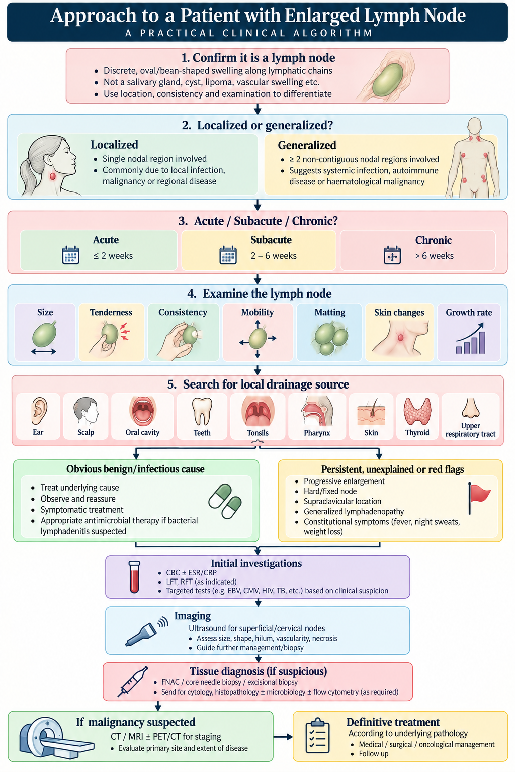
MANAGEMENT
Principle: treat the cause, not the lymph node itself.
Reactive/viral lymphadenopathy
Usually self-limiting. Needs
- Observation
- Reassurance
- Treatment of the underlying infection
- Analgesia/antipyretics if required
- Follow-up.
Acute bacterial lymphadenitis
Typical clinical picture: acute + tender + erythematous/warm + fever ± unilateral enlargement
Management may include:
- Appropriate antimicrobial therapy directed against likely organisms
- Analgesia
- Close reassessment.
Failure to improve should prompt reassessment for:
- Abscess
- Resistant organisms
- Atypical infection
- TB/NTM
- Malignancy
- Non-infectious causes.
- Ultrasound/CT confirmation
- Antibiotics
- Needle aspiration or surgical drainage depending on site and clinical circumstances.
Tuberculous lymphadenitis
Requires:
- Appropriate microbiological/histopathological confirmation where feasible
- Anti-tubercular therapy according to the applicable national/WHO regimen
- Assessment for pulmonary/extrapulmonary disease.
Surgical intervention is generally reserved for selected diagnostic or complicated situations rather than being the routine primary treatment.
Non-tuberculous mycobacterial lymphadenitis
Especially in children, classically presents as a slowly enlarging, relatively painless node, sometimes with overlying violaceous skin.
Options include:
- Observation in selected cases
- Antimicrobial therapy
- Surgical excision in selected cases.
Malignancy
Management depends on the diagnosis:
- Lymphoma → haematology/oncology-directed chemotherapy ± radiotherapy/targeted therapy.
- Metastatic carcinoma → identify primary tumour and stage disease; treatment may involve surgery, radiotherapy, systemic therapy or combinations.
- Leukemia → systemic hematological treatment.
Biopsy should generally precede definitive cancer-directed treatment, unless a specific emergency dictates otherwise.
- Gaddey & Riegel. Unexplained Lymphadenopathy: Evaluation and Differential Diagnosis. American Family Physician.
- Mohseni et al. Peripheral Lymphadenopathy: Approach and Diagnostic Tools. Iran J Med Sci.
- An approach to cervical lymphadenopathy in children. Singapore Medical Journal.
- Etiologies of Pediatric Cervical Lymphadenopathy: A Systematic Review of 2687 Subjects.
- Cervical: Lymphadenopathy - cervical nodal level classification and anatomy.
- Cervical lymphadenopathy: what radiologists need to know.