Open Academy navigation
Thyroid gland - Surgical anatomy and Physiology.
Introduction
Highly vascular endocrine organ situated in the anterior neck.
Regulated predominantly through the hypothalamic-pituitary-thyroid (HPT) axis
Cell type | Location | Hormone secreted | Hormone function |
|---|---|---|---|
Follicular cells (Thyrocytes) | Form the epithelial lining of thyroid follicles, surrounding the colloid | Thyroxine(T4) and Triiodothyronine (T3) | Regulate basal metabolic activity, thermogenesis, cardiovascular function, growth, skeletal maturation and neurological development |
Parafollicular cells (C-cells) | Located between follicular cells and in the interfollicular tissue; do not form the follicular lining | Calcitonin | Participates in calcium homeostasis, primarily by inhibiting osteoclastic bone resorption and thereby reducing serum calcium |
ANATOMY
Situation and extent
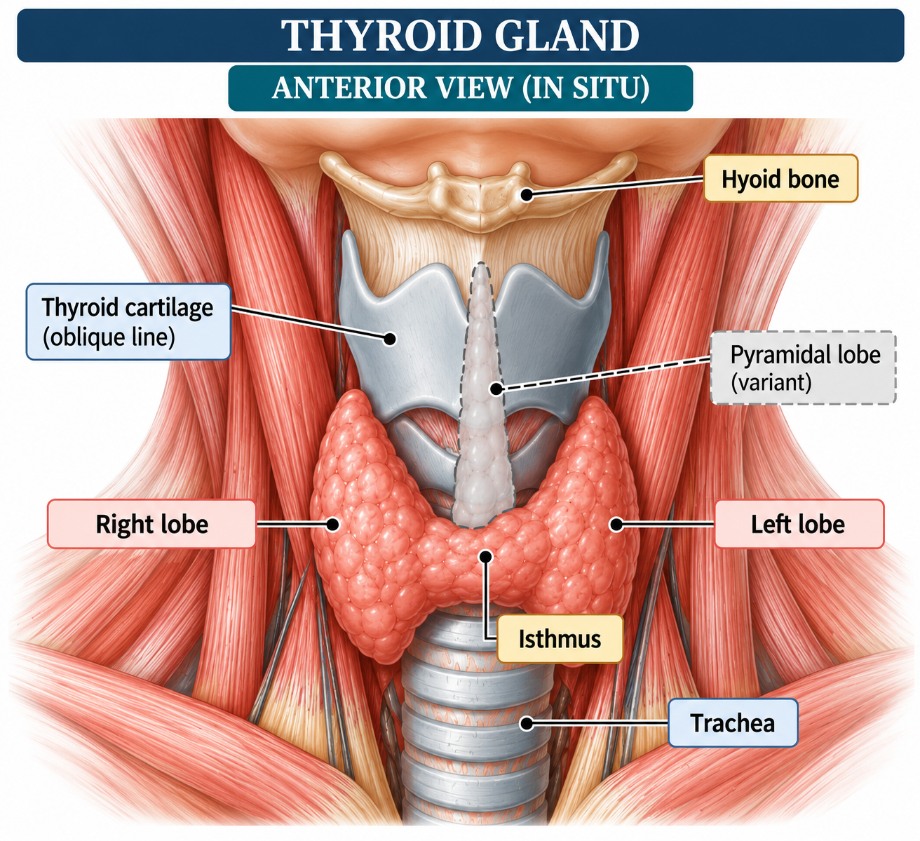
- Located in the anterior and lower part of the neck.
- Extends approximately from the level of the oblique line of the thyroid cartilage superiorly to the fifth or sixth tracheal ring inferiorly.
- Lies anterior and anterolateral to the larynx and upper trachea.
- weighs 15-25 g in adults (considerable physiological and pathological variation)
- Consists of:
- Right lobe
- Left lobe
- Isthmus
- A pyramidal lobe may arise from the isthmus or adjacent part of either lobe as a remnant of the thyroglossal duct. Pyramidal lobes have been reported in a substantial proportion of individuals and are clinically relevant during thyroidectomy.
External morphology
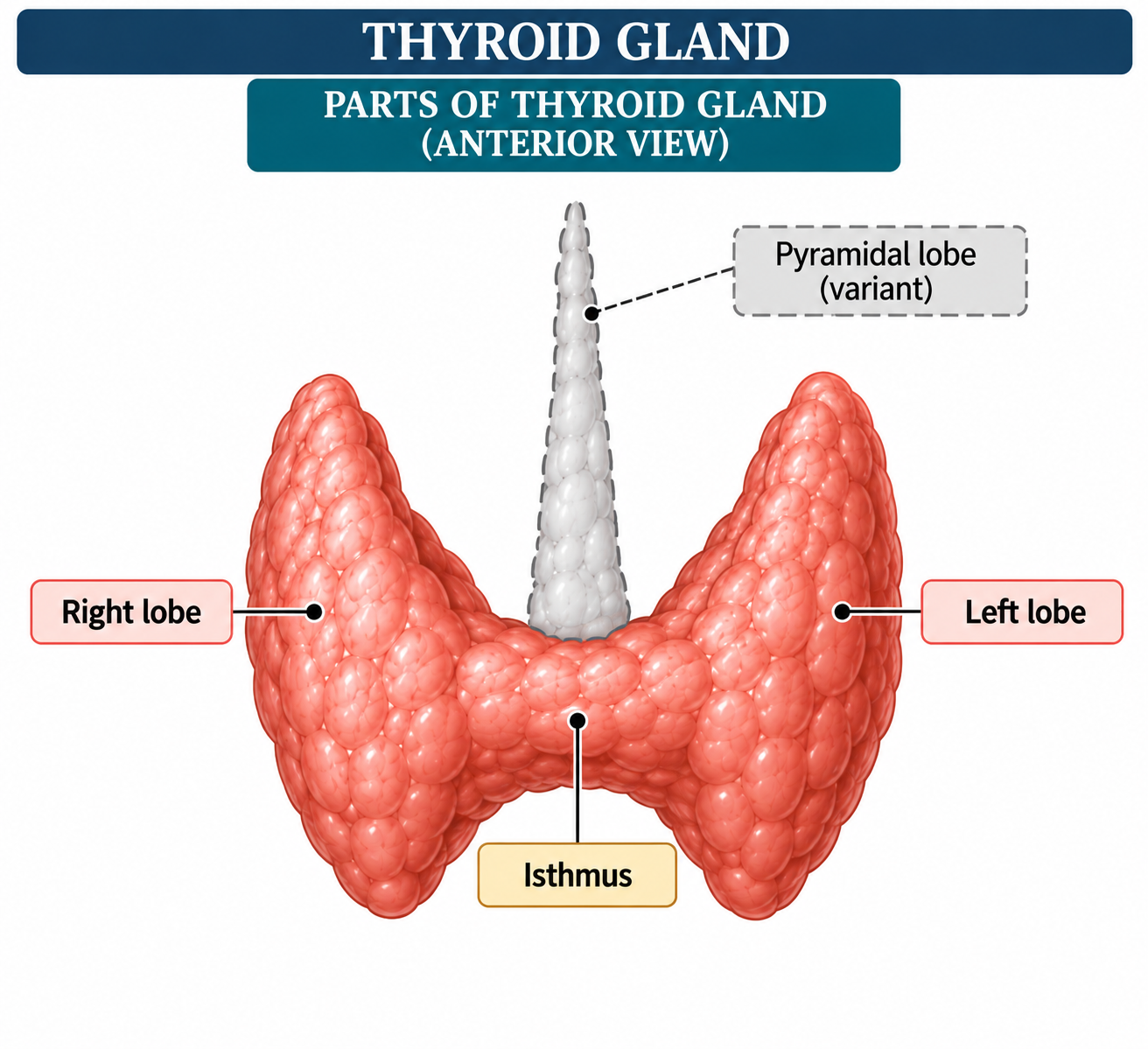
Each thyroid lobe is approximately conical, with:
- Apex directed superiorly.
- Base directed inferiorly.
- Anterolateral surface.
- Posteromedial surface.
- Medial surface related to the larynx, trachea and pharyngo-oesophageal structures.
- Posterior border related to the parathyroid glands and inferior thyroid vessels.
The two lobes are connected by the isthmus, which usually lies anterior to the second and third tracheal rings.
The gland is highly vascular and has a characteristic brownish-red appearance.
Capsules and fascial relations
The thyroid is surrounded by:
- A true fibrous capsule closely adherent to the gland.
- A false capsule derived from the pretracheal layer of deep cervical fascia.
The posterior condensation of the fascial attachment forms the ligament of Berry, which anchors the thyroid to the cricoid cartilage and upper tracheal rings.
The ligament of Berry is of major surgical importance because the recurrent laryngeal nerve is closely related to it near its entry into the larynx.
Relations
Anterior relations
- Skin
- Superficial fascia
- Platysma
- Investing fascia
- Infrahyoid muscles:
- Sternohyoid
- Sternothyroid
- Omohyoid
The sternothyroid muscle lies particularly close to the thyroid and may partially obscure the gland.
Posteromedial relations
- Larynx
- Trachea
- Pharynx
- Oesophagus
- Cricopharyngeus
- Inferior constrictor
- Recurrent laryngeal nerve
The recurrent laryngeal nerve ascends in close proximity to the tracheo-oesophageal groove and enters the larynx posterior to the cricothyroid joint.
Posterior relations
- Superior and inferior parathyroid glands
- Inferior thyroid artery and its branches
- Recurrent laryngeal nerve
- Sympathetic trunk lies posterior and medial to the carotid sheath rather than directly on the gland.
Lateral relations
- Carotid sheath
- Common carotid artery
- Internal jugular vein
- Vagus nerve
- Sternocleidomastoid muscle more superficially.
The close relationship of the thyroid to the carotid sheath, parathyroid glands and laryngeal nerves accounts for the importance of detailed surgical anatomy during thyroidectomy.
Microscopic structure
The thyroid is composed of numerous spherical follicles, which constitute the structural and functional units of the gland.
Each follicle consists of:
- A single layer of thyroid follicular epithelial cells.
- A central lumen containing colloid.
- A surrounding network of fenestrated capillaries.
The colloid consists predominantly of thyroglobulin, the protein matrix used for thyroid hormone synthesis.
Follicular cells are polarized:
- Basolateral surface → faces the bloodstream.
- Apical surface → faces the follicular lumen.
Parafollicular cells or C cells are located between follicular cells and within the interfollicular tissue. They secrete calcitonin.
Embryological significance
The thyroid develops primarily from an endodermal epithelial thickening in the floor of the primitive pharynx.
- It descends from the foramen caecum through the thyroglossal duct.
- The thyroglossal duct normally disappears.
- Persistence of its distal portion may produce a pyramidal lobe.
- Persistence of portions of the duct may result in thyroglossal duct cysts or fistulae.
- Ectopic thyroid tissue may occur anywhere along the embryological descent pathway.
The embryological course explains important anatomical variants encountered clinically and surgically.
VASCULAR SUPPLY
Arterial supply
The thyroid has an exceptionally rich arterial supply.
Major arteries:
- Superior thyroid artery
- Inferior thyroid artery
- Thyroid ima artery - occasional
Superior thyroid artery
- Usually the first anterior branch of the external carotid artery.
- Descends towards the superior pole of the thyroid.
- Supplies the upper part of the gland.
- Gives anterior and posterior glandular branches.
- Closely related to the external branch of the superior laryngeal nerve.
The origin of the superior thyroid artery is variable; it may arise from the external carotid artery, common carotid artery or carotid bifurcation.
Inferior thyroid artery
- Usually arises from the thyrocervical trunk of the subclavian artery.
- Courses medially and posteriorly behind the carotid sheath.
- Turns towards the posterolateral aspect of the thyroid.
- Supplies the inferior and posterior portions of the gland.
- Gives branches to the parathyroid glands.
Its terminal branches have an important relationship with the recurrent laryngeal nerve.
The recurrent laryngeal nerve may pass:
- Posterior to the inferior thyroid artery.
- Anterior to the artery.
- Between its branches.
Therefore, the relationship is variable and should not be used as the sole landmark for identifying the nerve.
Thyroid ima artery
An additional thyroid ima artery may occasionally be present.
- Usually ascends anteriorly from the brachiocephalic trunk or aortic arch.
- Reaches the inferior surface of the isthmus.
- May become clinically important during tracheostomy and lower cervical surgery.
- It may compensate for absence or hypoplasia of an inferior thyroid artery.
It is uncommon but should be considered during operative procedures.
Arterial anastomoses
The superior and inferior thyroid arteries form extensive anastomotic networks:
- Within the gland.
- Along the anterior and posterior surfaces.
- Between superior and inferior thyroid arterial territories.
- With neighbouring laryngeal and tracheal vessels.
This extensive vascularity explains the rapid haemorrhage that may occur following thyroid injury or surgery.
Venous drainage
A rich thyroid venous plexus lies on the surface and around the gland.
Three principal pairs of veins are described:
- Superior thyroid veins → internal jugular veins.
- Middle thyroid veins → internal jugular veins.
- Inferior thyroid veins → brachiocephalic veins.
An additional vein of Kocher may occasionally be present.
The inferior thyroid veins show greater anatomical variation than the superior and middle thyroid veins.
Lymphatic drainage
Lymphatic vessels form a dense network within the thyroid.
Drainage occurs primarily to:
- Prelaryngeal nodes.
- Pretracheal nodes.
- Paratracheal nodes.
- Upper and lower deep cervical lymph nodes.
Lymphatic spread is particularly important in thyroid malignancy, especially differentiated thyroid carcinoma.
NEURAL CONNECTIONS
Autonomic innervation
The thyroid receives autonomic fibres from:
- Sympathetic fibres from the cervical sympathetic chain.
- Parasympathetic fibres associated with the vagus nerve.
These fibres accompany the thyroid arteries and form a perivascular neural network.
The autonomic nerves primarily influence the vascular component of the gland rather than directly controlling thyroid hormone secretion. Thyroid hormone synthesis and secretion are principally regulated by TSH.
Recurrent laryngeal nerve
The recurrent laryngeal nerve is a branch of the vagus nerve.
Right side:
- Loops around the right subclavian artery.
- Ascends in the neck.
- Usually courses near the tracheo-oesophageal groove.
- Enters the larynx posterior to the cricothyroid joint.
Left side:
- Loops around the arch of the aorta near the ligamentum arteriosum.
- Ascends through the superior mediastinum.
- Continues in the tracheo-oesophageal groove.
- Enters the larynx posterior to the cricothyroid joint.
The nerve supplies:
- Motor innervation to all intrinsic laryngeal muscles except cricothyroid.
- Sensory innervation to the mucosa below the vocal cords.
- Sensory branches to the trachea and oesophagus.
Injury can result in:
- Hoarseness.
- Vocal cord paralysis.
- Ineffective cough.
- Dysphagia.
- Aspiration.
- Bilateral injury → potentially severe airway compromise.
The relationship between the recurrent laryngeal nerve and inferior thyroid artery is highly variable and therefore requires direct identification during thyroid surgery.
External branch of superior laryngeal nerve
The external branch of the superior laryngeal nerve:
- Arises from the superior laryngeal nerve, a branch of the vagus.
- Descends towards the superior thyroid pole.
- Runs close to the superior thyroid vessels.
- Supplies the cricothyroid muscle.
The cricothyroid muscle increases tension and length of the vocal folds and is important for high-pitched phonation.
Injury may produce:
- Difficulty producing high-pitched sounds.
- Vocal fatigue.
- Reduced voice projection.
- Subtle changes particularly important in professional voice users.
Individual ligation of the superior thyroid vessels close to the thyroid capsule helps reduce the risk of injury.
PHYSIOLOGY
Hypothalamic-pituitary-thyroid axis
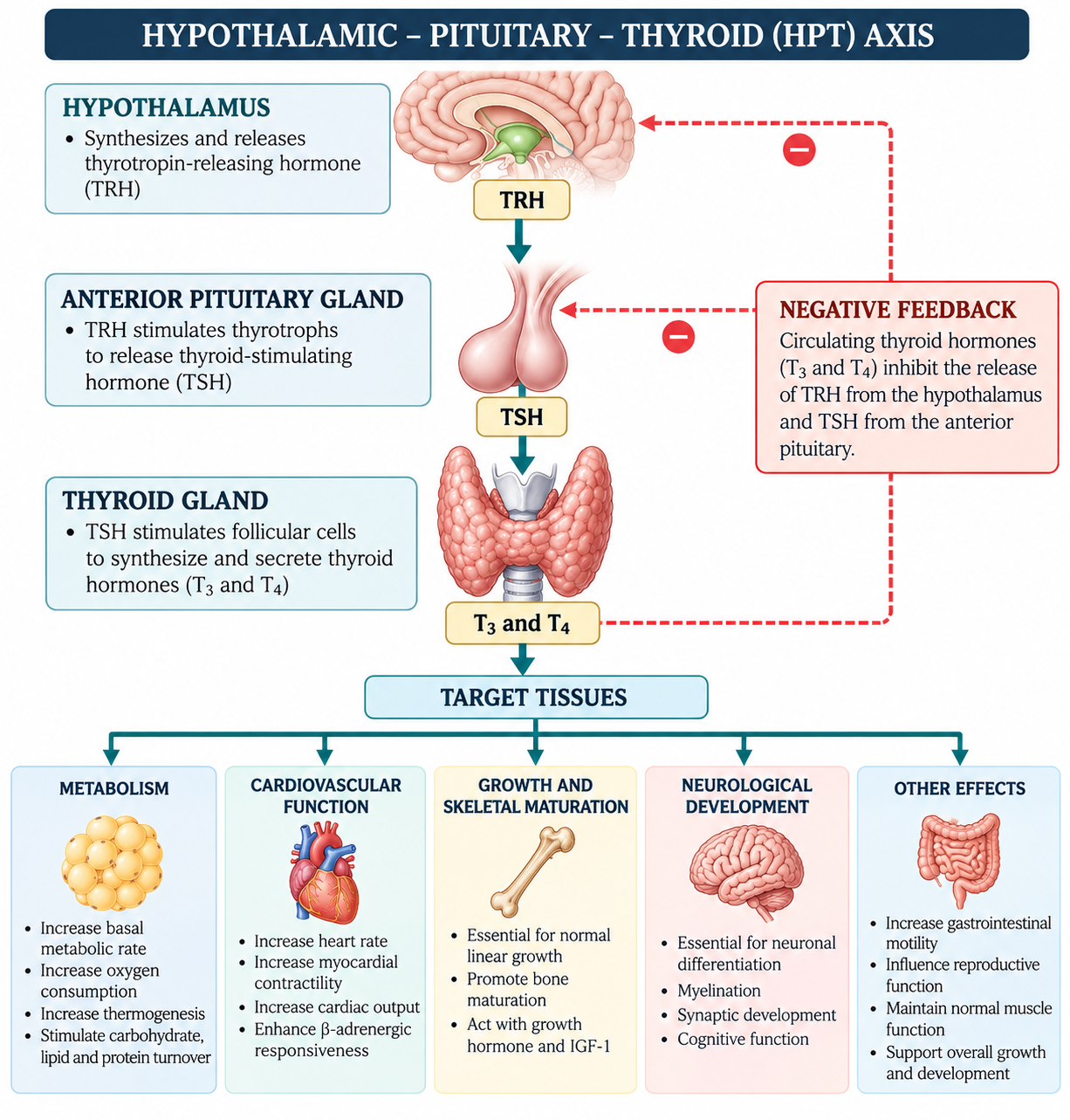
The HPT axis maintains circulating thyroid hormone concentrations within an appropriate physiological range.
- TRH is produced mainly by hypothalamic neurons.
- TRH stimulates thyrotrophs of the anterior pituitary.
- TSH stimulates thyroid follicular cells.
- TSH promotes thyroid hormone synthesis and secretion.
- Circulating T3 and T4 exert negative feedback on hypothalamus and pituitary.
This feedback maintains endocrine homeostasis.
Thyroid hormone synthesis
The major steps are:
- Iodide trapping: Iodide is actively transported into follicular cells through the sodium-iodide symporter.
- Transport to colloid: Iodide is transported across the apical membrane into the follicular lumen.
- Oxidation: Iodide is oxidized to reactive iodine by thyroid peroxidase (TPO).
- Organification: Iodine is attached to tyrosine residues within thyroglobulin.
- Formation of:
- MIT - monoiodotyrosine.
- DIT - diiodotyrosine.
- Formation of:
- Coupling
- MIT + DIT → T3.
- DIT + DIT → T4.
- Storage: T3 and T4 remain stored within thyroglobulin in the colloid.
- Endocytosis and proteolysis: Thyroglobulin is taken back into follicular cells. Lysosomal proteolysis releases T3 and T4.
- Secretion: T4 and T3 enter the circulation.
TSH stimulates several steps in this process, including iodide uptake, thyroglobulin synthesis and thyroid hormone secretion.
Peripheral conversion of thyroid hormones
The thyroid predominantly secretes T4. It is converted in peripheral tissues to:
- T3 - biologically more active.
- Reverse T3 - biologically inactive.
Important enzymes:
- Type 1 deiodinase.
- Type 2 deiodinase.
- Type 3 deiodinase.
T3 binds thyroid hormone receptors within target cells and regulates gene transcription.
Transport in blood
Most circulating thyroid hormone is protein-bound.
Major transport proteins:
- Thyroxine-binding globulin.
- Transthyretin.
- Albumin.
Only a small free fraction is biologically available to tissues.
Mechanism of thyroid hormone action
T3 enters target cells through specific transporters and binds nuclear thyroid hormone receptors.
The hormone-receptor complex:
- Interacts with thyroid hormone response elements.
- Alters gene transcription.
- Modifies protein synthesis.
- Produces tissue-specific metabolic and developmental effects.
Thyroid hormone also has important non-genomic actions involving intracellular signalling pathways.
PHYSIOLOGICAL FUNCTIONS
Metabolic functions
Thyroid hormones:
- Increase basal metabolic rate.
- Increase oxygen consumption.
- Increase mitochondrial activity.
- Stimulate carbohydrate utilization.
- Promote lipid turnover.
- Influence protein synthesis and degradation.
- Increase thermogenesis.
Thyroid hormone is therefore a major regulator of energy expenditure and oxidative metabolism.
Cardiovascular effects
Thyroid hormones:
- Increase heart rate.
- Increase myocardial contractility.
- Increase cardiac output.
- Enhance β-adrenergic responsiveness.
- Influence systemic vascular resistance.
Excess thyroid hormone may therefore produce tachycardia and increased cardiac workload, whereas deficiency may cause bradycardia and reduced cardiac output.
Growth and skeletal development
Thyroid hormones are essential for:
- Normal linear growth.
- Bone maturation.
- Epiphyseal development.
- Interaction with growth hormone and IGF-1.
- Normal skeletal development.
Severe thyroid hormone deficiency during childhood results in impaired growth and delayed skeletal maturation.
Brain and neurological development
Thyroid hormone is essential for:
- Neuronal differentiation.
- Myelination.
- Synaptic development.
- Normal cognitive development.
- Development of the central nervous system.
Thyroid hormone deficiency during critical periods of development can produce permanent neurological impairment if untreated.
Gastrointestinal functions
Thyroid hormones influence gastrointestinal motility.
- Excess → increased intestinal motility.
- Deficiency → reduced intestinal motility and constipation.
Musculoskeletal effects
Thyroid hormones influence:
- Muscle protein turnover.
- Muscle contraction.
- Bone remodelling.
- Skeletal maturation.
Both excess and deficiency can produce clinically significant neuromuscular manifestations.
Reproductive and endocrine effects
Normal thyroid function contributes to:
- Normal reproductive physiology.
- Menstrual regularity.
- Fertility.
- Pregnancy physiology.
- Interaction with other endocrine axes.
Thyroid dysfunction can alter reproductive function through effects on metabolism, gonadal function and hypothalamic-pituitary regulation.
CALCITONIN
Calcitonin is secreted by parafollicular C cells.
Major actions:
- Reduces osteoclastic bone resorption.
- Produces a modest reduction in serum calcium.
- Has a role in calcium homeostasis, although it is not essential for day-to-day calcium regulation in healthy adults.
Clinically, serum calcitonin is also used as a tumour marker in medullary thyroid carcinoma.
PATHOLOGICAL CONDITIONS
Thyroid disorders can be broadly classified into:
- Developmental abnormalities.
- Goitrous disorders.
- Functional disorders.
- Inflammatory disorders.
- Nodular disorders.
- Neoplastic disorders.
Developmental disorders
Thyroglossal duct cyst
- Results from persistence of the thyroglossal duct.
- Usually presents as a midline neck swelling.
- Often moves with swallowing and tongue protrusion.
- May become infected.
Ectopic thyroid
Possible sites include:
- Lingual thyroid.
- Sublingual region.
- Along the thyroglossal tract.
The ectopic tissue may represent the patient’s only functioning thyroid tissue.
Goitre
Goitre denotes enlargement of the thyroid gland.
Types include:
- Diffuse goitre.
- Multinodular goitre.
- Toxic goitre.
- Nontoxic goitre.
- Retrosternal/substernal goitre.
Causes include:
- Iodine deficiency or excess.
- Autoimmune disease.
- Dyshormonogenesis.
- TSH stimulation.
- Nodular thyroid disease.
Large goitres may produce compressive symptoms:
- Dysphagia.
- Dyspnoea.
- Stridor.
- Hoarseness.
- Venous compression.
Hyperthyroidism and thyrotoxicosis
Thyrotoxicosis refers to the clinical state produced by excessive circulating thyroid hormone.
Common causes:
- Graves disease.
- Toxic multinodular goitre.
- Toxic adenoma.
- Thyroiditis.
- Excessive exogenous thyroid hormone.
Typical manifestations:
- Weight loss.
- Heat intolerance.
- Sweating.
- Palpitations.
- Tremor.
- Anxiety.
- Increased bowel frequency.
- Menstrual disturbances.
- Tachycardia.
Graves disease
Graves disease is an autoimmune disorder in which thyroid-stimulating antibodies activate the TSH receptor.
Consequences:
- Diffuse thyroid enlargement.
- Increased thyroid hormone synthesis.
- Suppressed serum TSH.
- Elevated circulating free T4 and/or T3.
Extrathyroidal manifestations may include:
- Graves orbitopathy.
- Pretibial myxoedema.
Hypothyroidism
Hypothyroidism results from inadequate thyroid hormone action.
Common causes:
- Hashimoto thyroiditis.
- Iodine deficiency.
- Post-thyroidectomy state.
- Radioiodine therapy.
- Certain medications.
- Congenital thyroid disorders.
- Pituitary or hypothalamic disease.
Clinical manifestations:
- Fatigue.
- Cold intolerance.
- Weight gain.
- Constipation.
- Dry skin.
- Bradycardia.
- Reduced exercise tolerance.
- Cognitive slowing.
- Menstrual disturbances.
Hashimoto thyroiditis
Hashimoto thyroiditis is a chronic autoimmune thyroid disease characterized by immune-mediated destruction of thyroid follicles.
Histological features include:
- Dense lymphocytic infiltration.
- Lymphoid follicles with germinal centres.
- Follicular destruction and atrophy.
- Variable fibrosis.
- Hürthle cell change.
It commonly progresses to primary hypothyroidism.
Subacute thyroiditis
Subacute granulomatous thyroiditis is an inflammatory disorder that may follow a viral or post-viral illness.
Typical features:
- Painful thyroid enlargement.
- Fever or systemic symptoms.
- Elevated inflammatory markers.
- Transient thyrotoxicosis due to release of preformed hormone.
- Possible subsequent hypothyroid phase.
- Usually eventual recovery of thyroid function.
Histologically, granulomatous inflammation and multinucleated giant cells may occur.
Thyroid nodules
Thyroid nodules are common and may be:
- Cystic.
- Solid.
- Mixed.
- Benign.
- Malignant.
Important benign lesions include:
- Hyperplastic nodules.
- Colloid nodules.
- Follicular adenoma.
Evaluation commonly involves:
- Clinical examination.
- Thyroid function tests.
- Ultrasonography.
- Risk stratification according to sonographic features.
- Fine-needle aspiration cytology when indicated.
Thyroid malignancies
Major thyroid cancers include:
- Papillary thyroid carcinoma.
- Follicular thyroid carcinoma.
- Medullary thyroid carcinoma.
- Anaplastic thyroid carcinoma.
Papillary thyroid carcinoma
- Most common differentiated thyroid carcinoma.
- Frequently spreads through lymphatics to cervical lymph nodes.
- Characteristic nuclear changes are seen histologically.
- Generally arises from follicular epithelial cells.
Follicular thyroid carcinoma
- Arises from follicular cells.
- Characteristically spreads through vascular channels.
- Capsular and/or vascular invasion is important for diagnosis.
Medullary thyroid carcinoma
- Arises from parafollicular C cells.
- Produces calcitonin.
- May occur sporadically or as part of inherited syndromes such as MEN2.
The tubercle of Zuckerkandl is notable because of the high concentration of C cells in this region and therefore has surgical relevance in medullary thyroid carcinoma.
Anaplastic thyroid carcinoma
- Highly aggressive thyroid malignancy.
- Usually occurs in older adults.
- Presents with rapidly enlarging neck mass.
- May produce dysphagia, dyspnoea and airway compromise.
- Frequently invades surrounding structures.
CLINICAL AND SURGICAL IMPORTANCE
The thyroid gland is surrounded by structures essential for respiration, phonation, swallowing and calcium homeostasis.
Important structures to identify and preserve during thyroid surgery:
- Recurrent laryngeal nerve.
- External branch of superior laryngeal nerve.
- Superior and inferior parathyroid glands.
- Inferior thyroid artery and its terminal branches.
- Superior thyroid vessels.
- Thyroid venous plexus.
- Trachea.
- Oesophagus.
- Carotid sheath.
Major complications of thyroidectomy related to anatomy
- Haemorrhage and postoperative neck haematoma.
- Recurrent laryngeal nerve injury.
- External branch of superior laryngeal nerve injury.
- Hypoparathyroidism.
- Hypocalcaemia.
- Tracheal injury.
- Oesophageal injury.
- Injury to major cervical vessels.
The recurrent laryngeal nerve is particularly vulnerable near the ligament of Berry and its laryngeal entry point. The variable relationship between the nerve and inferior thyroid artery reinforces the importance of direct nerve identification rather than reliance on vascular landmarks alone.
SUMMARY
The thyroid gland is a highly vascular endocrine organ whose anatomical relationships have major physiological and surgical significance.
- Structurally, it consists of right and left lobes connected by an isthmus and may possess a pyramidal lobe.
- Microscopically, thyroid follicles containing colloid form the basic functional units.
- Its arterial supply is principally derived from the superior and inferior thyroid arteries, with an occasional thyroid ima artery.
- Venous drainage occurs through superior, middle and inferior thyroid veins.
- Autonomic fibres accompany the vascular supply, while the recurrent and superior laryngeal nerves have critical surgical relationships with the gland.
- Thyroid hormones regulate metabolic activity, thermogenesis, cardiovascular function, growth, skeletal maturation and neurological development.
- Thyroid activity is controlled predominantly by the hypothalamic-pituitary-thyroid axis.
- Major pathological conditions include goitre, thyroiditis, hyperthyroidism, hypothyroidism, thyroid nodules and thyroid malignancies.
- Detailed understanding of thyroid anatomy is essential for safe thyroid surgery because of the close relationship of the gland to the laryngeal nerves, parathyroid glands, trachea, oesophagus and major vessels.
REFERENCES
- Bhargav PR. Salient anatomical landmarks of thyroid and their practical significance in thyroid surgery: a pictorial review of thyroid surgical anatomy (revisited). Indian J Surg. 2014;76(3):207-211. doi:10.1007/s12262-013-0856-x. (PubMed Central (PMC))
- Barczynski M, et al. The anatomical relationship of inferior thyroid artery and recurrent laryngeal nerve: a review of the literature and its clinical importance. Int J Surg. 2020. (PubMed Central (PMC))
- Aytac B, et al. A review of methods for the preservation of laryngeal nerves during thyroidectomy. Int J Surg. 2020. (PubMed Central (PMC))
- Won HR, et al. Electrophysiological neural monitoring of the laryngeal nerves in thyroid surgery: review of the current literature. Clin Exp Otorhinol. 2015;8:1-7. (PubMed Central (PMC))
- Pujol M, et al. The thyroid gland: a revision study on its vascularization and surgical implications. Medicina (Kaunas). 2022;58. (PubMed Central (PMC))
- Natsis K, et al. Variations of venous drainage of the thyroid gland and their surgical implications: a narrative review. Surg Radiol Anat. 2023. (PubMed Central (PMC))
- Fekete C, Lechan RM. Central regulation of hypothalamic-pituitary-thyroid axis under physiological and pathophysiological conditions. Endocr Rev. 2014. (PubMed Central (PMC))
- Mullur R, Liu YY, Brent GA. Thyroid hormone regulation of metabolism. Physiol Rev. 2014;94:355-382. (PubMed Central (PMC))
- Brent GA. Mechanisms of thyroid hormone action. J Clin Invest. 2012;122:3035-3043. (PubMed Central (PMC))
- Schweizer U, et al. Molecules important for thyroid hormone synthesis and action-known facts and future perspectives. Endocr Rev. 2011. (PubMed Central (PMC))
- De Vito C, et al. Homeostatic control of the thyroid-pituitary axis: perspectives for diagnosis and treatment. Front Endocrinol. 2015. (PubMed Central (PMC))
- Korevaar TIM, et al. The thyroid hormone axis and female reproduction. Endocr Rev. 2023. (PubMed Central (PMC))