Open Academy navigation
Pashanagardabha (Parotitis) – Etiopathogenesis, Clinical features, Investigations, Differential Diagnosis, Complications and Management.
- Largest of the major salivary glands
- Predominantly serous gland.
- Occupies parotid bed
- Relations:
- Superiorly → external acoustic meatus and temporomandibular joint
- Inferiorly → angle of mandible and upper sternocleidomastoid region
- Anteriorly → masseter and mandibular ramus
- Posteriorly → mastoid process and sternocleidomastoid
- Medially → styloid apparatus, parapharyngeal structures and carotid space
- Enclosed by the parotid fascia, derived from the investing layer of deep cervical fascia.
Note: The parotid capsule is relatively firm. Therefore, acute inflammatory swelling can produce significant pain because the inflamed gland expands within a relatively confined fascial envelope.
LOBES
The facial nerve traverses the parotid & surgically divides into
- Superficial lobe
- Deep lobe
IMPORTANT STRUCTURES
- Facial nerve
- External carotid artery
- Retromandibular vein - Superficial temporal vein & Maxillary vein merge
- Intraparotid lymph nodes
- Auriculotemporal nerve branches
PAROTID DUCT - STENSEN DUCT
Parotid gland → crosses masseter → turns medially → pierces buccinator → opens into oral cavity opposite the upper second molar.
Examination: Massage the gland posterior to anterior. Observe the duct opening. Normal secretion is clear. Purulent discharge suggests bacterial sialadenitis.
- External carotid artery
- Superficial temporal artery
- Maxillary artery
- Transverse facial artery
- Retromandibular vein
NERVE SUPPLY
- Preganglionic fibres: CN IX → lesser petrosal nerve → otic ganglion
- Postganglionic fibres travel with the auriculotemporal nerve → parotid gland
- Auriculotemporal nerve
- Great auricular nerve
Facial nerve: passes through the gland but does not provide its secretomotor supply.
Intraparotid lymph nodes drain:
- Scalp
- External ear
- Periauricular region
- Eyelids
- Adjacent facial structures
Note:
- Reactive intraparotid lymphadenopathy is common.
- Metastatic cutaneous malignancies may involve the parotid.
- Lymphoma can arise within the parotid.
- Intraparotid nodes must be distinguished from primary parotid lesions.
ETIOPATHOGENESIS
Bacterial
- Staphylococcus aureus
- Streptococci
- Oral anaerobes
- Gram-negative organisms, particularly in hospitalised/immunocompromised patients
Viral
- Mumps
- Influenza
- Parainfluenza
- Coxsackievirus
- EBV
- CMV
- HIV
- Sialolithiasis
- Ductal stricture
- Mucus plug
- Ductal foreign body
- External compression
- Sjögren disease
- IgG4-related disease
- Sarcoidosis
- Granulomatosis with polyangiitis
- Other systemic inflammatory disorders
- Diabetes
- Malnutrition
- Liver disease
- Alcohol-associated disease
- Eating disorders
- Anticholinergic drugs
- Radiation
- Radioiodine
- Certain medications causing xerostomia
- Postoperative dehydration
- Pneumoparotitis
- Positive-pressure ventilation
- Wind-instrument playing
G. Neoplastic/pseudoneoplastic
- Pleomorphic adenoma
- Warthin tumour
- Mucoepidermoid carcinoma
- Adenoid cystic carcinoma
- Lymphoma
- Metastatic disease
Reduced salivary flow may occur with:
- Dehydration
- Fever
- Fasting
- Postoperative state
- Anticholinergic drugs
- Poor oral intake
- Malnutrition
- Severe systemic illness
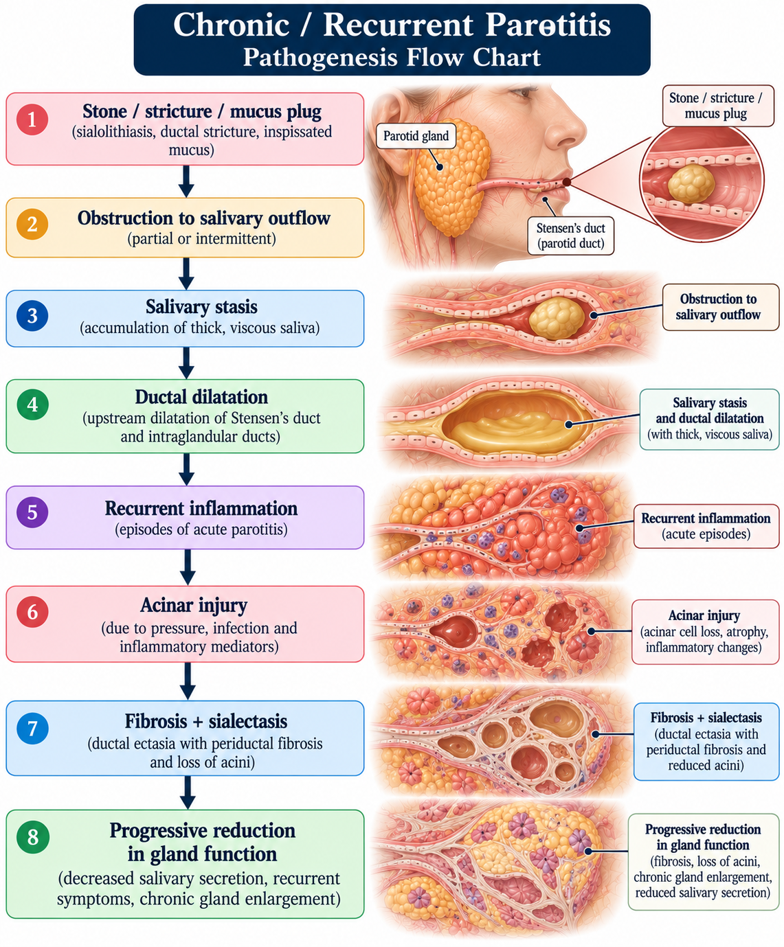
OBSTRUCTION → SALIVARY STASIS → INFLAMMATION → INFECTION
Chronic obstructive parotitis becomes a self-perpetuating cycle. Hence repeated antibiotics fail in mechanical obstructive disease.
CLINICAL CLASSIFICATION
Pattern | Important possibilities |
|---|---|
Acute + painful + unilateral | Bacterial parotitis, obstruction with secondary infection |
Acute + painful + bilateral | Viral parotitis, systemic disease |
Recurrent + meal-related pain | Sialolithiasis/ductal obstruction |
Chronic + bilateral | Sjögren, HIV, sarcoidosis, metabolic causes |
Chronic + painless + discrete mass | Benign tumour |
Painless + firm + persistent | Neoplasm/autoimmune disease |
Swelling + facial palsy | Malignancy until proven otherwise |
CLINICAL FEATURES
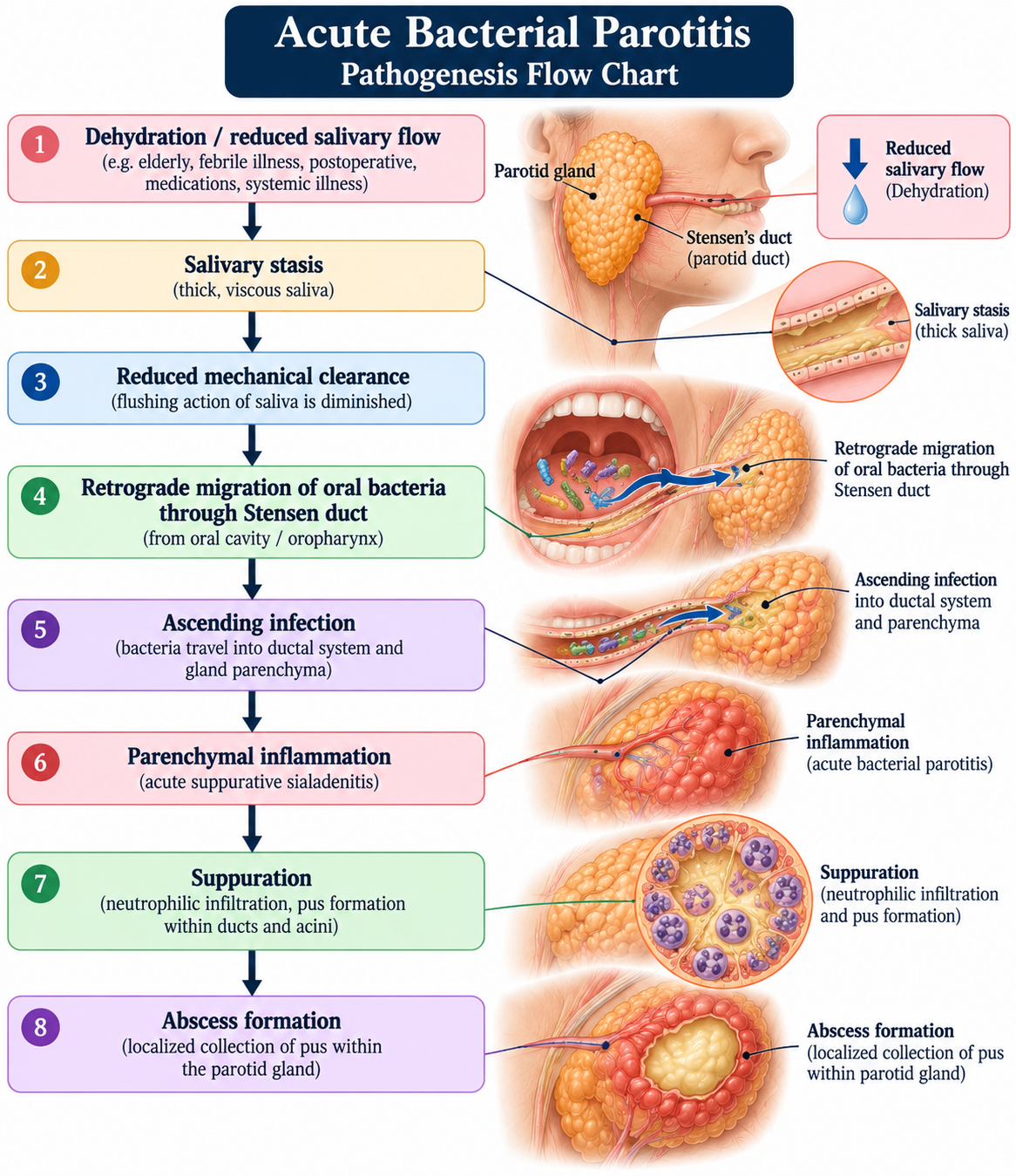
associated with dehydration, poor oral hygiene, debility and salivary stasis.
- Sudden painful swelling
- Usually unilateral
- Tender gland
- Fever
- Malaise
- Erythema
- Warmth
- Pain on chewing
- Trismus in more severe disease
- Reduced salivary flow
- Purulent discharge from Stensen duct
VIRAL PAROTITIS - MUMPS
- Fever
- Headache
- Malaise
- Myalgia
- Anorexia
- followed by painful swelling of one or both parotids - peaks within 1–3 days & resolves in a week. Unilateral in 25% patients.
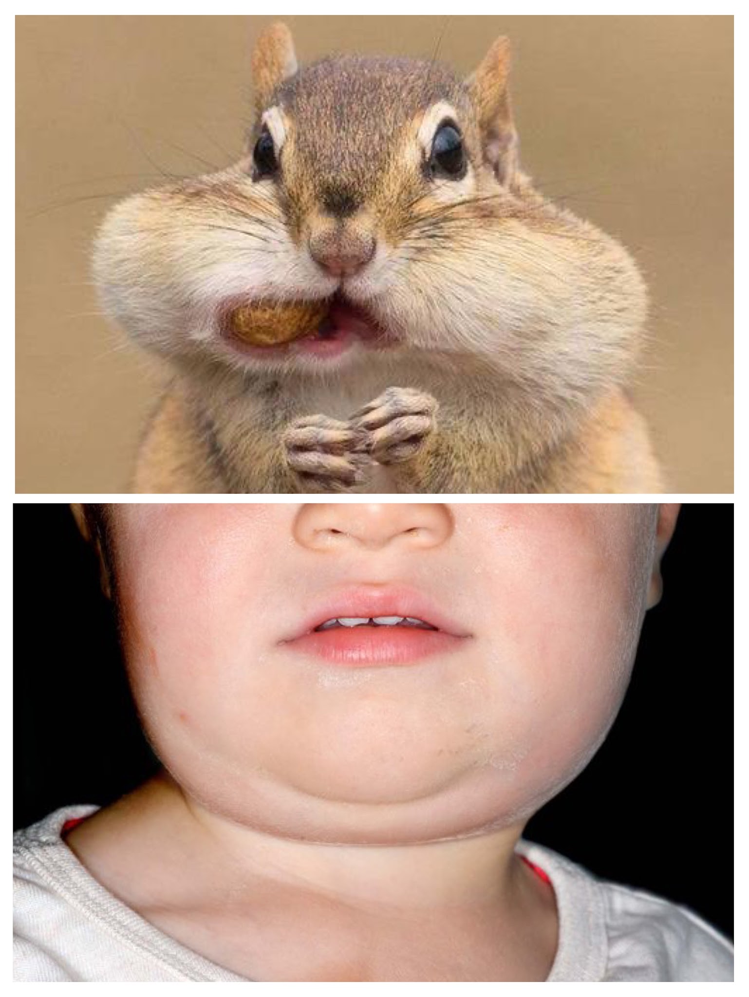
- Chipmunk Cheek / Puffy cheek appearance: angle of the mandible becomes obscured and the ear may be displaced upward and outward.
Mumps can occur even in vaccinated individuals, vaccination may reduce risk and severity.
OBSTRUCTIVE PAROTITIS - Meal-time syndrome
Food stimulus → increased salivary secretion → obstruction → painful glandular enlargement.
- Pain immediately before/during meals
- Recurrent swelling
- Gradual resolution after eating
- Recurrent episodes over months/years
A palpable stone may occasionally be identified - Sialolithiasis
CHRONIC PAROTITIS
Features include:
- Recurrent swelling
- Recurrent pain
- Reduced salivary secretion
- Thick/abnormal saliva
- Xerostomia
- Repeated infective episodes
- Progressive glandular fibrosis
Sialography: Fruit-laden tree / branchless fruit-laden tree appearance
SJÖGREN-ASSOCIATED PAROTITIS
- Bilateral parotid enlargement persistently
- Xerostomia
- Dry eyes
- Recurrent gland swelling
- Dental caries
- Difficulty swallowing dry foods
- Increased risk of B-cell lymphoma
EXAMINATION
- Unilateral/bilateral swelling
- Skin erythema
- Ear displacement
- Facial asymmetry
- Discrete mass
- Scars
- Fistula
- Temperature
- Tenderness
- Consistency
- Fluctuation
- Mobility
- Discrete versus diffuse enlargement
Massage the gland and observe duct opening.
- Clear saliva
- Thick saliva
- Purulent discharge
- Blood
- Absence of secretion
- CN VII - Facial weakness + parotid mass - malignant involvement.
- CN V
- CN IX/X
- CN XII
- Cervical lymph nodes
INVESTIGATIONS
CBC - Leukocytosis & Neutrophilia in bacterial disease.
- Gram stain + culture + sensitivity
- If recurrent/unusual
- Anaerobic culture
- Mycobacterial studies
- Fungal studies
- If abscess: US-guided aspiration
ULTRASONOGRAPHY
- Non-invasive
- No radiation
- Easily available
- Can identify gland enlargement
- Detects duct dilatation
- Detects stones
- Detects abscess
- Identifies lymph nodes
- Guides aspiration/FNAC
- Differentiates diffuse inflammatory enlargement vs focal lesion.
- Suspected abscess
- Deep-space extension
- Facial/deep neck involvement
- Bony involvement
- Calculi
- Complications
- Ductal dilatation
- Fat stranding
MRI
- Deep-lobe lesions
- Tumours
- Perineural spread
- Soft-tissue extension
- Complex inflammatory disease
- Neoplasia
- Vascular lesion
CONTRAST SIALOGRAPHY
- Ductal stenosis
- Sialectasis
- Ductal obstruction
- Chronic inflammatory changes
Note: Acute suppurative infection is a contraindication - ductal instrumentation exacerbates inflammatory process.
- Direct visualisation of duct
- Identification of stones
- Strictures
- Mucus plugs
- Inflammatory changes
- Stone removal
- Duct dilation
- Irrigation
- Removal of debris
- Intraductal medication
Major modern development in salivary gland surgery. This has shifted the treatment paradigm toward minimally invasive, gland-preserving management for selected obstructive/recurrent disease.
Consider when there is:
- Persistent focal mass
- Suspicion of tumour
- Lymphadenopathy
- Persistent unilateral enlargement
- Atypical chronic disease
FNAC is particularly useful for differentiating inflammatory enlargement from neoplastic disease, although the choice between FNAC and core biopsy depends on the clinical/imaging scenario.
AUTOIMMUNE INVESTIGATIONS
When Sjögren’s or systemic autoimmune disease is suspected:
- ANA
- Anti-Ro/SSA
- Anti-La/SSB
- Rheumatoid factor
- Immunoglobulins
Additional investigations should be guided by the clinical phenotype.
INFECTIOUS WORKUP
Depending on epidemiology and clinical suspicion:
- Buccal swab RT-PCR
- Viral testing
- Serology in appropriate circumstances
CDC recommends laboratory testing when mumps is suspected; RT-PCR from buccal/oral specimens is an important diagnostic test.
Consider particularly with:
- Bilateral chronic parotid enlargement
- Cystic lesions
- Xerostomia
- Generalised lymphadenopathy
- Recurrent unexplained parotitis
Consider in endemic settings with:
- Chronic unilateral swelling
- Cervical lymphadenopathy
- Constitutional symptoms
- Granulomatous pathology
DIFFERENTIAL DIAGNOSIS
- Acute bacterial parotitis
- Mumps
- EBV
- CMV
- Influenza
- HIV-associated salivary disease
- TB
- Other viral infections
- Sialolithiasis
- Ductal stricture
- Mucus plug
- Ductal foreign body
- Sjögren disease
- IgG4-related disease
- Sarcoidosis
- Granulomatosis with polyangiitis
- Sialadenosis
- Diabetes
- Alcohol-associated enlargement
- Malnutrition
- Liver disease
- Eating disorders
- Pleomorphic adenoma
- Warthin tumour
- Oncocytoma
- Other benign salivary tumours
- Mucoepidermoid carcinoma
- Adenoid cystic carcinoma
- Acinic cell carcinoma
- Salivary duct carcinoma
- Carcinoma ex pleomorphic adenoma
- Lymphoma
- Metastatic cutaneous malignancy
Feature | Bacterial | Viral/mumps | Obstructive | Autoimmune | Tumour |
|---|---|---|---|---|---|
Onset | Acute | Acute | Recurrent | Chronic/recurrent | Gradual |
Pain | Marked | Moderate | Meal-related | Variable | Usually absent |
Fever | Common | Common | Usually absent unless infected | Usually absent | Usually absent |
Laterality | Usually unilateral | Often bilateral | Usually unilateral | Often bilateral | Usually unilateral |
Pus from duct | Often | No | Usually no | Usually no | No |
Meal association | Variable | No | Strong | Variable | No |
Discrete mass | Usually no | No | Sometimes | Usually no | Common |
Facial palsy | Complication | Unusual | Unusual | Unusual | Major warning sign |
Imaging | Inflammation/abscess | Diffuse enlargement | Stone/stricture | Diffuse heterogeneity | Focal lesion |
MANAGEMENT
Identify & treat the cause: Infection/ Obstruction/ Autoimmune/ Neoplastic/ Metabolic
ACUTE BACTERIAL PAROTITIS
- Massage
- Antibiotics
- Sialogogues - Stimulate salivary flow
- Hydration
- Analgesia
- Warm compresses
- Oral hygiene
- Correction of predisposing factors
Clinical clues: Severe focal tenderness, Fluctuation, Persistent fever, Failure to improve with antibiotics, Progressive swelling, Trismus & Imaging showing collection
- Antibiotics
- Imaging
- Culture
- Ultrasound-guided aspiration
- Drainage
OBSTRUCTIVE PAROTITIS
- Hydration
- Gland massage
- Sialogogues
- Analgesia
- Treatment of secondary infection
- Small ductal stone → endoscopic removal where feasible.
- Stricture → sialendoscopic dilatation.
- Larger/complex stone → endoscopy-assisted or combined endoscopic–external approaches.
CHRONIC/RECURRENT PAROTITIS
- Hydration
- Oral hygiene
- Massage
- Sialogogues
- Analgesics
- Sialendoscopy
- Ductal lavage
- Stricture dilatation
- Stone removal
Refractory(stubborn/irresponsive) disease- Parotidectomy
For Sjögren-associated disease:
- Salivary stimulation
- Hydration
- Dental care
- Management of xerostomia
- Treatment of systemic autoimmune disease
- Immunomodulatory therapy in selected patients
Persistent/ suspicious gland enlargement - ? lymphoma.
MUMPS PAROTITIS
No specific antiviral therapy if uncomplicated.
- Rest
- Hydration
- Analgesia/antipyretics
- Nutritional support
- Isolation
SURGICAL MANAGEMENT:
The shift from gland excision toward minimally invasive gland preservation is an important development in modern salivary surgery.
Surgical Caution - Facial nerve injury/ Haemorrhage from RM vein & EC artery/ Fistula/ Scar/ Incomplete drainage - Hence imaging-guided intervention & knowledge of parotid surgical anatomy are important.
Abscess → aspiration and/or drainage depending on location, size and response.
Persistent obstruction → sialendoscopy / stone extraction / ductal intervention.
Recurrent chronic → gland-preserving procedures where possible; else parotidectomy.
Tumour → appropriate oncologic parotid surgery.
Cellulitis - surrounding soft tissues.
- Facial nerve palsy
Systemic Complications (In Mumps)
- Orchitis
- Oophoritis
- Mastitis
- Pancreatitis
- Meningitis
- Encephalitis
- Hearing loss
Post-surgical Complications
- Facial nerve injury → temporary or permanent facial weakness.
- Frey syndrome - Gustatory sweating due to aberrant regeneration of parasympathetic fibres after parotid surgery.
- Salivary fistula
- Sialocele
- Haemorrhage - RM vein/EC artery.
- Infection
- Cosmetic deformity
- First-bite syndrome - particularly deep parotid/parapharyngeal surgeries.
- Persistent unilateral swelling : tumour / chronic obstruction / granulomatous disease
- Discrete firm mass: neoplasm
- Facial nerve palsy : major malignant warning sign until evaluated.
- Cervical/intraparotid lymphadenopathy: Malignancy/ Lymphoma/ Infection
- Bilateral persistent enlargement: Sjögren’s/ HIV/ Sarcoidosis/ IgG4-related disease/ Sialosis
- Recurrent meal-related pain: ductal obstruction / sialolithiasis
- Failure to improve with antibiotics: Abscess/ Obstruction/ Resistant organism/ Autoimmune disease/ Malignancy
- Al Ghabra Y, Goldin J, Pandey S. Parotitis. StatPearls. Updated June 23, 2025. NCBI Bookshelf.
- Wilson KF, Meier JD, Ward PD. Salivary gland disorders. American Family Physician. 2014;89:882–888.
- Schmal F, Stoll W, et al. Approach to sialadenitis. Canadian Family Physician. 2023;69:757–762.
- Raad II, Sabbagh MF, Caranasos GJ. Acute bacterial sialadenitis: a study of 29 cases and review. Reviews of Infectious Diseases. 1990;12:591–601.
- Brook I. Acute bacterial suppurative parotitis: microbiology and management. Journal of Cranio-Maxillofacial Surgery / related review literature.
- Spiegel JH, et al. Diagnosis and management of parotitis. Review of infectious, obstructive and inflammatory causes.
- Zenk J, Iro H, Klintworth N, Lell M. Diagnostic imaging in sialadenitis. Oral and Maxillofacial Surgery Clinics of North America. 2009;21:275–292.
- Ji YD, Donoff RB, Peacock ZS, Carlson ER. Surgical Landmarks to Locating the Main Trunk of the Facial Nerve in Parotid Surgery: A Systematic Review. Journal of Oral and Maxillofacial Surgery. 2018;76:438–443.
- Pather N, Osman M. Landmarks of the facial nerve: implications for parotidectomy. Surgical and Radiologic Anatomy. 2006;28:170–175.
- de Ru JA, van Benthem PPG, Bleys RLAW, Lubsen H, Hordijk GJ. Landmarks for parotid gland surgery. Journal of Laryngology & Otology. 2001;115:122–125.
- Rea PM, McGarry G, Shaw-Dunn J. The precision of four commonly used surgical landmarks for locating the facial nerve in anterograde parotidectomy in humans. Annals of Anatomy. 2010;192:27–32.
- Saibene AM, Allevi F, Ayad T, et al. Treatment for parotid abscess: a systematic review. Acta Otorhinolaryngologica Italica. 2022;42:106–115.
- Tan VES, Goh BS. Parotid abscess: a five-year review-clinical presentation, diagnosis and management. Journal of Laryngology & Otology. 2007;121:872–879.
- Centers for Disease Control and Prevention. Clinical Features of Mumps. Updated June 5, 2024.
- Centers for Disease Control and Prevention. Mumps: Pink Book-Pathogenesis, Clinical Features, Laboratory Testing and Complications.
- Centers for Disease Control and Prevention. Clinical Overview of Mumps. Updated January 17, 2025.
- Review of the Major and Minor Salivary Glands, Part 1: Anatomy, Infectious, and Inflammatory Processes. Radiologic/clinical review literature.
- Bilateral parotid swelling: a radiological review. Insights into Imaging.
- Parotid gland sialolithiasis: a comprehensive systematic review and meta-analysis. Recent systematic review of gland-preserving and endoscopic treatment approaches.
हनुसन्धौ समुद्भूतं शोफमल्परुजं स्थिरम् ।
पाषाणगर्दभं विद्यात् कफवातसमन्वितम् ॥
Correlation: Parotitis (inflammation of the parotid gland)
- स्थान : हनुसन्धि - Temporomandibular joint
- स्वरूप : स्थिर, कठिन पाषाणवत् शोफ (firm, stone-like swelling).
- वेदना : अल्प (mild pain).
- दोष : कफ-वात
- निरुक्ति : पाषाणवत् काठिन्य (stone-like hardness).
- लेप - with paste of मनःशिला, हरताल, कुष्ठ & दारु
- पाचन - Allow swelling to mature.
- भेदन - Incision when fully ripened.
- व्रणवत् उपचार - Manage subsequently like a wound.
References:
- सुश्रुत संहिता, निदानस्थान, अध्याय 13 - क्षुद्ररोग निदान
- सुश्रुत संहिता, चिकित्सास्थान, अध्याय 18 - क्षुद्ररोग चिकित्सा